Monday, August 29, 2016

steatocystoma multiplex

Steatocystoma multiplex:
Definition:
It is also called as epidermal poly cystic disease.
There are numerous epithelial lined sebum filled dermal cysts with characteristic sebaceous gland in the cystic  wall.

Associated with Alagille syndrome and pachyonychia congenita type 2.
In these cases defect is K17 mutations.
It can be sporadic occurrence or inherited as autosomal dominant.

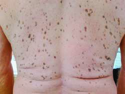
Clinical presentation:
Skin coloured cysts on trunk, upper arms, scrotum and chest.

No symptoms
Complication: suppurations and sinus formation leading to scarring.

When single cyst is present in sporadic manner  it's called steatocystoma simplex.

Pathological findings:
Cyst is lined by stratified squamous epithelium but no granular layer  and sebaceous glands are located on cyst wall.
Eosinophilic cuticle on luminal side of this wall with hair and keratin inside it.

Differential diagnosis: eruptive vellus hair cyst
                                        Epidermal inclusion cyst

Treatment:
Surgical excision
Cyst drainage with manual removal of cyst.
Inflamed lesions can be treated with oral retinoids, cryotherapy or CO2 laser or intralesional steroids.


Monday, August 15, 2016

Lasers

Therapeutic applications of lasers:

1. Tattoo removal

2. Vascular lesions:
A. Telangiectasia
B. Port wine stain
C. Infantile haemangioma
E. Venous lake
F. Starburst veins
G. Varicose veins
H. Lymphangioma circumscripta
I.  Tuberous sclerosis ( angiofibromas)


3. Tumors:
A. Trichoepithelioma
B. Neurofibromas
C. digital mucous cyst
D. Xanthelesma
E. Syringomas.                                      

4. Nevi:
A. Epidermal nevus
B. Sebaceous nevi
C. Congenital melanocytic nevus
D. Nevus of Ota
E. Nevus zygomaticus
F. Nevus pigmentosus

5. Hair removal
6. Alopecia areata
7. Vitiligo
8. Keloid and hypertrophic scars
9. Acne and acne scars
10. Seborrhoeic keratoses
11. Freckles
12. Solar lentigo
13. Skin resurfacing 


Monday, August 8, 2016

Biological therapy in dermatology

Rituximab 375mg/m2 weekly for 4weeks in pemphigus
Mechanism of action for Rituximab:
It is anti CD20 monoclonal antibody.


Biological agents in psoriasis:
1) Ustekinumab:
45 mg SC on week 0,4, then every 12 weeks

2) Secukinumab:Initial: 300 mg SC at weeks 0, 1, 2, 3, and 4
Monthly maintenance: Beginning at week 8, give 300 mg SC once monthly
For some patients, a dose of 150 mg may be acceptable

3) Adalimumab: 80mg SC on day 1
                        40mg on day 8
                        40 mg every 2 weeks
These agents can be combined with phototherapy or methotrexate.
 Ustekinumab has shown maximum disease free period after treatment.
Remember:
All biological agents are found to be less effective in obese patients when used as a monotherapy.

Adalimumab can be used in hidradenitis suppurativa cases.

First generation biological agents for psoriasis: etarncept, Adalimumab, inflixiamb
These agents target TNF -alpha.
Second generation: Ustekinumab acts against IL-12/23( pro- inflammatory cytokines)
Third generation: targets IL-17.

Tuesday, August 2, 2016

Seborrheic keratoses

Seborrheic keratoses (SK) :
It's a benign epidermal tumour.
It is commonly present in old age.
It's types:
1) Common seborrheic keratoses
2) Skin tags
3) Irritant SK
4) Pigmented SK
5) SK with squamous atypia
6) Stucco keratoses
7) Leser trelat sign
8) Reticulated SK
9) Dermatosis papulosa Nigra (DPN)
10) Clonal SK

Histopathology:
Acanthosis, papillomatosis, pseudo cyst formation, hyper keratosis.

Clinical features:
Stuck on appearance of lesion

Treatment:
Cryotherapy
Electric desiccation
Laser
Surgical excision
Topical flurouracil

Biopsy is recommended when
Rapid growth of lesion
Atypical clinical appearance
Symptomatology
Unusual lesion location
In the above features there is risk of malignant transformation.

Irritant SK may develop in patients who are on chemotherapy especially cytarabine drug.

Lesar Trelat sign:
Eruption of SK on trunk in Christmas tree pattern
Cause: underlying adenocarcinoma of stomach most commonly
Other: adenocarcinoma of lung and colon.

DPN :
Present in skin type 4 and above.

    Fig: seborrhoeic keratoses 

Fig:Leser trelat sign

Tattoo removal

Tattoo removal:
Tattoos are developed  with  different colours.
Tattoo made by India ink or graphite are easy to remove.
But tattoos made by green,yellow and orange inks are resistant to laser treatment.
Black tattoos show good response to laser therapy.

Methods to remove tattoo:
1) Deep cryotherapy
2) CO2 laser vaporisation
3) Salabrasion
4) Dermabrasion
5) Topical agents
These methods can cause scarring as side effect.

Lasers are good option but recurrence or incomplete removal do happen.
Mechanics behind laser:
Pigment in dermal phagocytes is exposed to temp upto 300 degrees in nanoseconds. This leads to rupture of cell and alteration of chemicals.

Remember:
Red tattoo should be treated by green laser (Q-switched Nd YAG) 532 nm
Green tattoo must be cleared by red colour laser ( ruby 694 nm) or alexandrite laser.
Different colours need to be treated with appropriate corresponding wavelengths of laser.

Multiple sessions of laser are required.
Immediate side effect:
1) Immediate whitening of skin over tattoo is end point of laser therapy.
This will disappear few minutes after cessation of laser treatment as gas bubbles of laser settle down.
2) Epidermal pigment cells also suffer in laser treatment so transient hypo pigmentation occurs.

The risk of scarring from Q- switched laser treatments is approximately 5 percent.

Monday, August 1, 2016

Intolerance to cosmetics


1) Examine every cosmetic and skin-care product
2) Administer patch and photopatch tests to rule out occult
allergic and photoallergic dermatitis
3) Test for contact urticaria
4) Do careful repeat open application testing (ROAT)
5) Treat endogenous inflammatory disease
6) Limit skin-care to:
Water washing without soap or detergent
Lip cosmetics as desired, if lips are clear
Eye cosmetics as desired, if eyelids are asymptomatic
Face powder
Glycerin and rose water as a moisturizer, if necessary.


Cosmetic intolerance syndrome:
It is extreme end of irritant or allergic contact dermatitis to cosmetic products.
Patient can not tolerate any cosmetics over face due to burning sensation.
Definition :
It is an uncommon clinical phenomenon in which patients complain of facial burning and discomfort associated with application of most skin-care products.

Severity of symptoms does not match objective signs of disease.

 status cosmeticus:  condition in which patients no longer tolerate the use of any facial cosmetic product.

 irritation to occult allergic contact dermatitis, allergic photocontact dermatitis, contact urticarial reaction, or a combination can be presenting symptoms.
Investigations:
careful clinical review and patch testing of casual agents.
Advice:
 prolonged program of eliminating cosmetics is  important.

dermatologic nondisease: facial burning continuously, without showing objective signs.