Tattoo removal:
Tattoos are developed with different colours.
Tattoo made by India ink or graphite are easy to remove.
But tattoos made by green,yellow and orange inks are resistant to laser treatment.
Black tattoos show good response to laser therapy.
Methods to remove tattoo:
1) Deep cryotherapy
2) CO2 laser vaporisation
3) Salabrasion
4) Dermabrasion
5) Topical agents
These methods can cause scarring as side effect.
Lasers are good option but recurrence or incomplete removal do happen.
Mechanics behind laser:
Pigment in dermal phagocytes is exposed to temp upto 300 degrees in nanoseconds. This leads to rupture of cell and alteration of chemicals.
Remember:
Red tattoo should be treated by green laser (Q-switched Nd YAG) 532 nm
Green tattoo must be cleared by red colour laser ( ruby 694 nm) or alexandrite laser.
Different colours need to be treated with appropriate corresponding wavelengths of laser.
Multiple sessions of laser are required.
Immediate side effect:
1) Immediate whitening of skin over tattoo is end point of laser therapy.
This will disappear few minutes after cessation of laser treatment as gas bubbles of laser settle down.
2) Epidermal pigment cells also suffer in laser treatment so transient hypo pigmentation occurs.
The risk of scarring from Q- switched laser treatments is approximately 5 percent.
Hi there, I am Dr. Snehal Thorat specialized in Dermatology and cosmetology. Presently taking advance training in lasers at China Medical University Hospital of Taiwan. If you have any skin related issues or about cosmetic problems, please contact me.
Tuesday, August 2, 2016
Monday, August 1, 2016
Intolerance to cosmetics
1) Examine every cosmetic and skin-care product
2) Administer patch and photopatch tests to rule out occult
allergic and photoallergic dermatitis
3) Test for contact urticaria
4) Do careful repeat open application testing (ROAT)
5) Treat endogenous inflammatory disease
6) Limit skin-care to:
Water washing without soap or detergent
Lip cosmetics as desired, if lips are clear
Eye cosmetics as desired, if eyelids are asymptomatic
Face powder
Glycerin and rose water as a moisturizer, if necessary.
Cosmetic intolerance syndrome:
It is extreme end of irritant or allergic contact dermatitis to cosmetic products.
Patient can not tolerate any cosmetics over face due to burning sensation.
Definition :
It is an uncommon clinical phenomenon in which patients complain of facial burning and discomfort associated with application of most skin-care products.
Severity of symptoms does not match objective signs of disease.
status cosmeticus: condition in which patients no longer tolerate the use of any facial cosmetic product.
irritation to occult allergic contact dermatitis, allergic photocontact dermatitis, contact urticarial reaction, or a combination can be presenting symptoms.
Investigations:
careful clinical review and patch testing of casual agents.
Advice:
prolonged program of eliminating cosmetics is important.
dermatologic nondisease: facial burning continuously, without showing objective signs.
Saturday, July 30, 2016
Aquagenic syringeal acrokearatoderma
Aquagenic syringeal acrokeratoderma:
It is eccrine gland disease.
Pathogenesis is poorly understood
Stratum corneum dysfunction and aberration in eccrine gland structure can be possible for the disease presentation.
Clinical features:
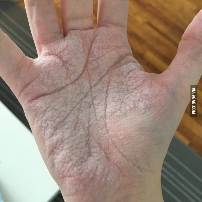
Patient complains of appearance of whitish translucent Papules over palms after immersion of hand in water.
Once the contact with water is removed it disappears in 10-20 minutes.
Sometimes it is accompanied by sensation of tingling and tightness of palm.
Excessive wrinkling of skin over palms develop on repeated exposures
Other sites:
Dorsal hands or fingers
Rarely sole is involved
Associations:
Cystic fibrosis
Family history (+) in some cases
Hyperhidrosis
Conditions should be distinguished from hereditary papulotranslucent acrokearatoderma.
Histopathology is helpful to confirm diagnosis
On histopathology orthokeratosis and dialated eccrine ducts are seen in aquagenic acrosyringeal kearatoderma while eccrine gland is normal in hereditary variant.
Treatment:
No standard treatment is available
Options:
1) Aluminum chloride hexahydrate 20% at night
2) Barrier emollients petroleum based
3) 12% ammonium lactate cream
4) Botulinum toxin injection
Biopsy should be performed immediately after immersion of hand in water.
This disease is rare but sometimes it may be under diagnosed.
The hand in bucket sign is very typical of this condition.
It is eccrine gland disease.
Pathogenesis is poorly understood
Stratum corneum dysfunction and aberration in eccrine gland structure can be possible for the disease presentation.
Clinical features:
Patient complains of appearance of whitish translucent Papules over palms after immersion of hand in water.
Once the contact with water is removed it disappears in 10-20 minutes.
Sometimes it is accompanied by sensation of tingling and tightness of palm.
Excessive wrinkling of skin over palms develop on repeated exposures
Other sites:
Dorsal hands or fingers
Rarely sole is involved
Associations:
Cystic fibrosis
Family history (+) in some cases
Hyperhidrosis
Conditions should be distinguished from hereditary papulotranslucent acrokearatoderma.
Histopathology is helpful to confirm diagnosis
On histopathology orthokeratosis and dialated eccrine ducts are seen in aquagenic acrosyringeal kearatoderma while eccrine gland is normal in hereditary variant.
Treatment:
No standard treatment is available
Options:
1) Aluminum chloride hexahydrate 20% at night
2) Barrier emollients petroleum based
3) 12% ammonium lactate cream
4) Botulinum toxin injection
Biopsy should be performed immediately after immersion of hand in water.
This disease is rare but sometimes it may be under diagnosed.
The hand in bucket sign is very typical of this condition.
Multiple whitish Papules over palm after water exposure
Thursday, July 28, 2016
Freckles
Freckles:
It is pigmentory condition.
It's also called ephelids.
Freckles are brownish pigmented Macules of various sizes over face.
Other sites: axilla and inguinal regions.
When these sites other than face are involved then we suspect neurofibromatosis type 1.
Freckles are also present in one genodermatoses called xeroderma pigmentosa.
Most patients have freckles only over face. In that case it's very easy to treat it.
Diagnosis is solely based on clinical examination.
Treatment:
1) Topical treatment:
A)Use of Depigmenting agents : Kojic acid
B) Phenol 80% over spots
2) lasers:
a)Frequency doubled Nd. YAG 532 nm
b) IPL (500-1000 nm )
c) Q switched ruby laser 694nm
d) 755nm alexandrite laser
e) 532 nm KTP laser
3)chemical peel
Side effects: post inflammatory hyper or hypo pigmentation.
Remember: freckles don't require treatment always.
When cosmetic concern is there treatment options can be chosen.
Freckles do have 2.5 fold increased risk of sunburn and poor response to tanning.
Difference between freckles and lentigo:
Freckles. Lentigo
1) Darkening on sun exposure No change
2) Presents in early childhood Elderly people
3) No increase in melanocyte Increase in melanocyte number
Number
Use of sunscreen with spf 30+ is strongly recommended in these patients.
It is pigmentory condition.
It's also called ephelids.
Freckles are brownish pigmented Macules of various sizes over face.
Other sites: axilla and inguinal regions.
When these sites other than face are involved then we suspect neurofibromatosis type 1.
Freckles are also present in one genodermatoses called xeroderma pigmentosa.
Most patients have freckles only over face. In that case it's very easy to treat it.
Diagnosis is solely based on clinical examination.
Treatment:
1) Topical treatment:
A)Use of Depigmenting agents : Kojic acid
B) Phenol 80% over spots
2) lasers:
a)Frequency doubled Nd. YAG 532 nm
b) IPL (500-1000 nm )
c) Q switched ruby laser 694nm
d) 755nm alexandrite laser
e) 532 nm KTP laser
3)chemical peel
Side effects: post inflammatory hyper or hypo pigmentation.
Remember: freckles don't require treatment always.
When cosmetic concern is there treatment options can be chosen.
Freckles do have 2.5 fold increased risk of sunburn and poor response to tanning.
Difference between freckles and lentigo:
Freckles. Lentigo
1) Darkening on sun exposure No change
2) Presents in early childhood Elderly people
3) No increase in melanocyte Increase in melanocyte number
Number
Use of sunscreen with spf 30+ is strongly recommended in these patients.
Tuesday, July 26, 2016
Moisturizer
Moisturizer:
It is to maintain moisture of your skin.
It is used very routinely by females.
But do you know which moisturizer is the best for you?
I will educate you about the mechanism behind moisturizer...

Types:
1) occlusive
2) humectant 3) emollient
It is to maintain moisture of your skin.
It is used very routinely by females.
But do you know which moisturizer is the best for you?
I will educate you about the mechanism behind moisturizer...
Types:
1) occlusive
2) humectant 3) emollient
Emollient = occlusive + humectant
occlusive:
Beeswax, soybean oil
Humectants: urea, sugar, sorbitol.
Emollient: lanolin
Beeswax, soybean oil
Humectants: urea, sugar, sorbitol.
Emollient: lanolin
Melasma
Melasma :
It is a pigmentory disorder characterised by brownish Macules over face especially cheeks and forehead.
Types:
1) Epidermal
2) Dermal
3) Mixed
Depending on the level of pigmentation into the skin.
Another classification is based on the location of skin affected
1) Malar
2) Mandibular
3) Centrofacial
Woods light is used to determine the type of melasma.
Cause:
1)Increased estrogen levels in females especially pregnancy
2) prolonged sun exposure
When it happens after or during pregnancy it's called chloasma (pregnancy mask).
How to treat??
1) Topical treatment
2) Systemic
3) Laser or light therapy
4) Chemical peel
1) Topical treatment:
A) Depigmenting agents:
1) Kojic acid
2) Mulberry extract
3) Hydroquinone 4%
B) Retinoids
C) Steroids
Most common topical treatment :
Kligman formula (Triple combination formula):
Hydroquinone+ tretinoin+ fluocinonide
1) Systemic treatment:
a) Capsule vitamin c 200mg 1OD for 2-3 months.
b) Tab tranexamic acid 250-500 mg 1OD
2) Lasers:
Nd Yag laser toning 1064 nm is good for maintenance.
Intense pulsed light therapy is another alternative.
3) Chemical peel:
a) Glycolic acid peel 30%
Peeling procedures have downtime so most of the patients don't prefer.
Mesotherapy with tranexamic acid is also tried in some centres.
Recurrence rate is high with any above treatment.
No standard treatment is available. It is all depends on individual skin type.
Advice :
1) Use of sunscreen with spf 50+ and UVA+++
2) Use of wide brimmed hat in mid-day sun
3) Use of retinoids at night time
It is a pigmentory disorder characterised by brownish Macules over face especially cheeks and forehead.
Types:
1) Epidermal
2) Dermal
3) Mixed
Depending on the level of pigmentation into the skin.
Another classification is based on the location of skin affected
1) Malar
2) Mandibular
3) Centrofacial
Woods light is used to determine the type of melasma.
Cause:
1)Increased estrogen levels in females especially pregnancy
2) prolonged sun exposure
When it happens after or during pregnancy it's called chloasma (pregnancy mask).
How to treat??
1) Topical treatment
2) Systemic
3) Laser or light therapy
4) Chemical peel
1) Topical treatment:
A) Depigmenting agents:
1) Kojic acid
2) Mulberry extract
3) Hydroquinone 4%
B) Retinoids
C) Steroids
Most common topical treatment :
Kligman formula (Triple combination formula):
Hydroquinone+ tretinoin+ fluocinonide
1) Systemic treatment:
a) Capsule vitamin c 200mg 1OD for 2-3 months.
b) Tab tranexamic acid 250-500 mg 1OD
2) Lasers:
Nd Yag laser toning 1064 nm is good for maintenance.
Intense pulsed light therapy is another alternative.
3) Chemical peel:
a) Glycolic acid peel 30%
Peeling procedures have downtime so most of the patients don't prefer.
Mesotherapy with tranexamic acid is also tried in some centres.
Recurrence rate is high with any above treatment.
No standard treatment is available. It is all depends on individual skin type.
Advice :
1) Use of sunscreen with spf 50+ and UVA+++
2) Use of wide brimmed hat in mid-day sun
3) Use of retinoids at night time
Monday, July 25, 2016
Skin types
Skin types :
Main four types:
1) dry vs oily
2) pigmented vs non pigmented
3) sensitive vs irritant
4) wrinkled vs tight
With these basic types individual skin can fall in any of 16 types
skin catogerization is important while treating any skin disease
It is important while choosing over the counter cosmetic products.
So the following table will help you to define your skin type.
Oily sensitive skin is more prone to acne and or rosacea.
Pigmented skin should be protected from excessive sun exposure.
Dry skin is likely to develop eczema.
Fitzpatrick skin typing is universal method
Subscribe to:
Posts (Atom)